MECA-OM
The Optical Microscope
|
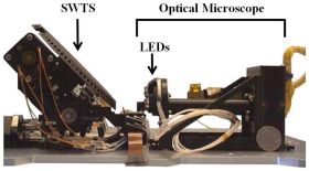
The MPS also provided the focal plane with the Charge Coupled Device (CCD) detector for the Optical Microscope (figure) that is part of the MECA (Microscopy and Electrical Conductivity Analyzer) payload.
|

Figure: The Optical Microscope with its sample holder (Sample Wheel Translation Stage, SWTS) and its Light Emitting Diodes (LEDs). The entire setup is (together with the MECA Wet Chemistry Cells) enclosed in a rectangular box on the lander deck. The SWTS can receive soil samples from the scoop. Then it is translated and rotated in order to bring the collected sample in the field of view of the microscope.
|
Science objectives
The Optical Microscope can receive soil material from the digging site via the scoop. In a first place this material is dropped on various targets (magnetic, non-magnetic, textured or sticky targets) of a particular sample holder, the so-called SWTS (Sample Wheel Translation Stage). Then the SWTS is translated and rotated such that the delivered material can be imaged by the Optical Microscope. These images provide information on color, grain size (down to < 10 microns), texture and porosity as well as magnetic properties. In some sense the Optical Microscope extends the RAC data down to the micrometer scale.
The Instrument
The Optical Microscope is a fixed-focus imaging system with 6x magnification and a resolution of 4 microns/px at the location of the target. It has the same CCD as the RAC. Both instruments share a common readout electronics that is located elsewhere on the lander (cf. MPS-RAC link for further information on the CCD).
The Optical Microscope has 4 types of light sources: Red, green, blue and UV Light Emitting Diodes (3 LEDs of each type). Acquiring images, while either red, green or blue LEDs are switched on, provides color information of the soil samples and allows for generation of true-color images of these samples.
The CCD detector is not sensitive in the spectral region (λ ~ 350 nm), where the UV LED emits. Sending UV light onto the target can thus reveal luminescence of that target. The amount of luminescence can then be assessed by comparison with the one from a UV calibration target that is also available on the SWTS.
Characteristics of the MECA-OM
| Magnification |
5.75 |
| CCD |
256 x 512 pixel |
| Imaged area |
1 mm x 2 mm |
| Resolution |
4 microns/px |
| Light source |
3 red, 3 green, 3 blue and 3 UV LEDs |
MPS Contribution
Focal plane with CCD
The Team
| Horst Uwe Keller (PI) |
| Wojtek Markiewicz |
| Stubbe F. Hviid |
| Rainer Kramm |
| Hermann Hartwig |
| Hermann Arnemann |
| Helmut Schüddekopf |
| Walter Goetz |
Related links
